
|
|
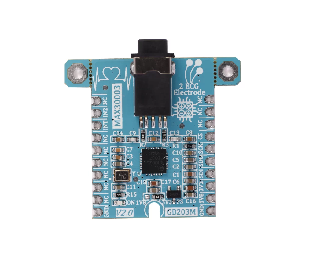
Product
|
Type
|
Resolution
|
Channel
|
Interface
|
Sampling Rate
|
Buy
|
DOC
|
|---|---|---|---|---|---|---|---|---|
|
Digital
|
16-Bit
|
2-channel
|
SPI
|
125 SPS to 8 kSPS
|
|
||
|
Digital
|
16-Bit
|
2-channel
|
SPI
|
125 SPS to 8 kSPS
|
|
||
|
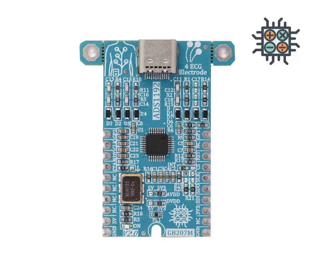
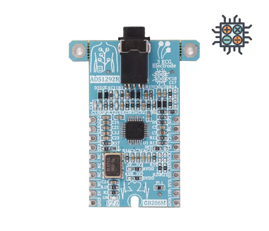
Digital
|
24-Bit
|
2-channel
|
SPI
|
125 SPS to 8 kSPS
|
|
||
|
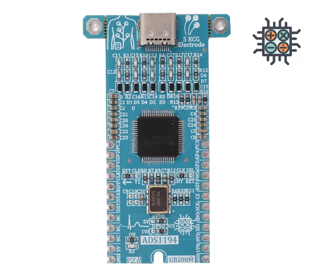
Digital
|
16-Bit
|
8-channel
|
SPI
|
125SPS to 8kSPS
|
|
||
|
Digital
|
18-Bit
|
single-channel
|
SPI
|
125 SPS to 512 SPS
|
|
||
|
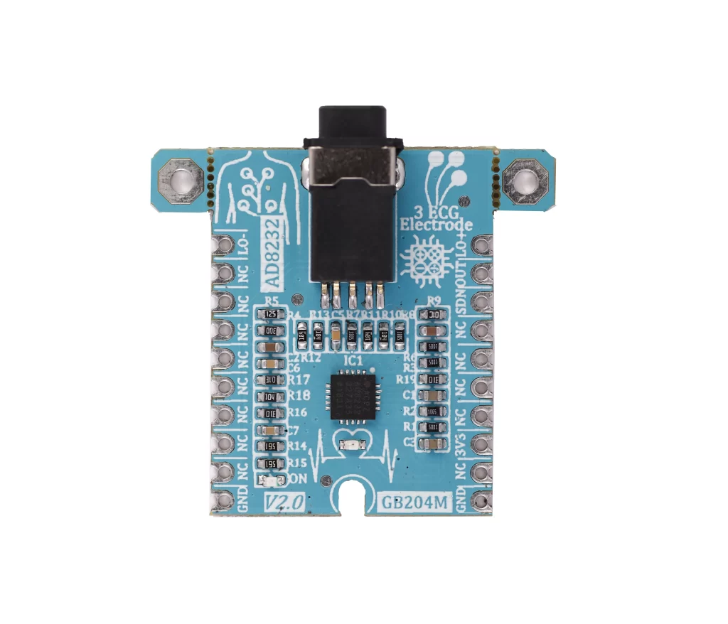
Analog
|
---
|
---
|
Analog
|
360SPS to 2KSPS
|
|